
Preparation of equipment
The different parts of the fermenter and their functions were studied and summarized in the table below:

Preparation of media

1. 50 g of Luria Bertani (LB) medium was dissolved in 2L of water in a 2L bottle.
2. 100mL of the LB broth was then transferred in a 200mL shaker flask and the remaining 1900mL was transferred in the 2-L bioreactor.
3. The media was autoclaved at 121°C for 20 minutes.

4. When the broth has cooled down below 50°C, ampicillin was added to both the seed and fermentation media to a final concentration of 100µg/mL.
5. The media was then kept at 4°C until inoculation.
Preparation of culture (all the steps in this protocol were done aseptically)

1. pGLO transformed E. coli was retrieved from the -80°C freezer.
2. The cells were then streaked on a LB/Amp/Ara plate in the following manner showed below:

3. The plate was then incubated for 24 hours.
4. On the following day, several colonies formed on the plate were transferred to the flask containing 100mL LB medium with ampicillin.
5. The flask was then placed in a shaking incubator to incubate at 32°C for 24 hours.
Setting up and Monitoring Fermentation Process

1. After autoclaving, the 1900mL media that was remaining in the 2L bottle was transferred to the bioreactor.
2. The control parameters were then set as shown below:

3. A sample of 2mL was taken from the bioreactor before inoculation.
4. 100mL of the prepared seed culture was then inoculated into the bioreactor and fermentation was continued for 24 hours at the above conditions.

5. Samples of 2mL were taken from the bioreactor every hour for a period of 10 hours.
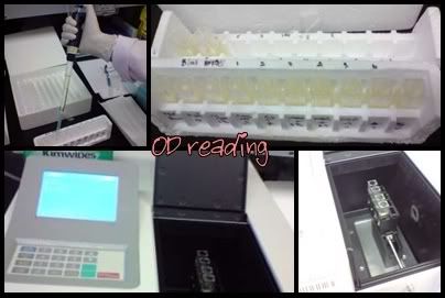
6. The optical density of the fractions were recorded at 600 nm
Isolation of GFP

· The cells were obtained by centrifugation at 10,000rpm for 5 minutes.
· The pellet was resuspended in 500µLof TE buffer of pH 7.5 and 2 drops of lysozyme were added.
· After 15 minutes, the tube was placed in liquid nitrogen till the contents were frozen.
· The tube was then thawed in warm water and the cycle of freezing and thawing was repeated for 2 times.
· Sonication was done on ice for 4 cycles of 25 seconds with 10 seconds rest in between sonication cycles.
· The contents of the tubes were spun down at 20 minutes at 10,000rpm
· The pellet and supernatant were separated and the pellet was resuspended using 400µL of TE buffer.
Purification of GFP

· 8 test tubes were labelled from 1-8 and one labelled blank.
· The blank was filled with 2.0mL of ammonium bicarbonate and the rest of the tubes were marked at the 2.0mL level.
· The column was carefully drained into a waste beaker.
· The cell free extract was transferred to the top of the gel bed
· The fractions were obtained by placing test tubes under the stopcock and filling each tube to a level of 2mL
· 50mM of ammonium bicarbonate were continuously added to the top of the column while taking the fractions.
· The optical density of the fractions were recorded again at 476 nm
Labels: procedure